
Advances in biological imaging have given scientists unprecedented datasets with extremely high resolutions, yet data interpretation tools are working overtime to keep up. This is particularly evident in the case of cryo-electron tomograms (cryo-ET), where the samples exhibit inherently low contrast due to the limited electron dose that can be applied during imaging before radiation damage occurs.
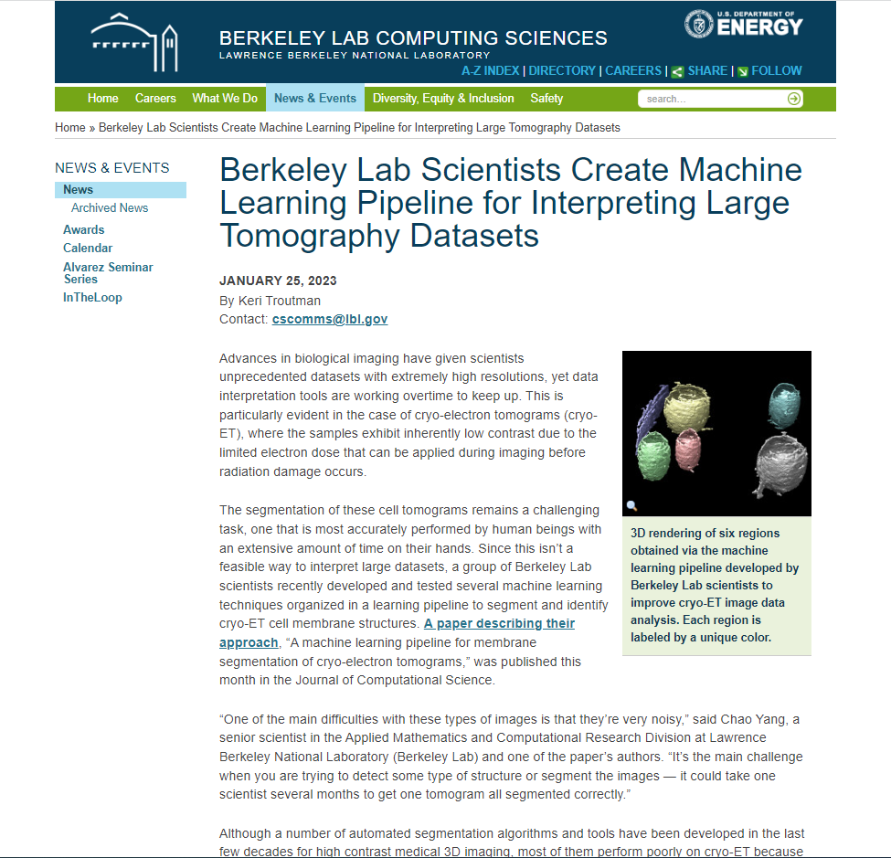
The segmentation of these cell tomograms remains a challenging task, one that is most accurately performed by human beings with an extensive amount of time on their hands. Since this isn’t a feasible way to interpret large datasets, a group of Berkeley Lab scientists recently developed and tested several machine learning techniques organized in a learning pipeline to segment and identify cryo-ET cell membrane structures. A paper describing their…


